Calcite and Sphalerite - Huanzala Mine, Bolognesi Province, Ancash, Peru
- M Crawford (Post by M Cole)
- Aug 10, 2021
- 2 min read
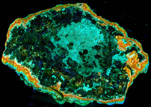
This is a multi-color longwave fluorescent rock from the Huanzala Mine, Bolognesi Province, Ancash, Peru. The specimen has red-orange fluorescent calcite crystals on top of yellow fluorescent sphalerite. The yellow fluorescent sphalerite covers a dark red fluorescent mineral. I sent chips of this mineral for XRD analysis and the results identified the mineral as sphalerite. This is the first time that I have seen red fluorescent sphalerite. Longwave UV spectra of this red sphalerite show that the fluorescent response is brighter in the near infrared. There are two broad peaks in the near infrared around 730nm and 870nm. This is the brightest and most broad near infrared response that I have encountered so far in my study of red and infrared fluorescent minerals.
Glenn Waychunas talked about red fluorescent sphalerite in the most recent Journal of the Fluorescent Mineral Society. A trace amount of copper can activate a red fluorescent response in sphalerite. Robbin’s book describes how iron can also activate red fluorescence in sphalerite. Not sure which of these activators or some other activator causes the red and near infrared fluorescence in this specimen. Manganese is the fluorescent activator of the calcite and the “yellow” sphalerite.
The pictures show the longwave UV fluorescence. The first picture is the true color image of the longwave UV fluorescence. The second picture is a color infrared image. This image is formed by assigning the near infrared image to red, red visible light to green and green visible light to blue. The bright red area in this color infrared image shows the near infrared fluorescence of the sphalerite. The calcite and the “yellow” sphalerite have no near infrared fluorescence and appear green and blue-green in the color infrared image. The third picture is the white light image of the specimen.
The fourth picture shows longwave UV spectra of the three fluorescent minerals. The last set of pictures shows the back of the specimen. The pictures are:
True color image of the longwave UV fluorescence.
A black and white image of the near infrared fluorescence caused by longwave UV illumination. The picture was taken with a 780nm filter. Wavelengths less than 780nm are blocked by the filter. Near infrared light with wavelengths longer than780nm are passed by the filter
A false color image of red=near infrared fluorescence, green=red visible fluorescence, blue=green visible fluorescence.
White light image of the backside of the specimen.