sodalite specimen from Sar-e-Sang, Badakhshan, Afghanistan
- M Crawford (Post by M Cole)
- Aug 8, 2021
- 2 min read
I like to collect fluorescent rocks that also tell a story about their formation. This is a sodalite specimen from Sar-e-Sang, Badakhshan, Afghanistan. This sodalite specimen has been brecciated and the voids were filled white dolomite. The dolomite identification is from XRD analysis. The dolomite also contains grains of light brown titanite/sphene.
The emplacement of the dolomite-titanite modified the visible and fluorescent properties of the sodalite. The original sodalite has a blue color. The fluids that deposited the dolomite and titanite altered the sodalite to a violet color. Several of blue sodalite clasts have a reaction rim of violet sodalite where they are in contact with the dolomite. Additionally, longwave UV fluorescent response of the altered sodalite is slightly different. The peak fluorescent response is shifted to longer wavelengths (see Picture 7). The altered sodalite became phosphorescent and tenebrescent. Titanium from the dolomite fluids penetrated into the sodalite clasts and is the likely activator for the phosphorescence. The reaction rims are clearly shown in the phosphorescent image (Picture 4). The emplacement of the dolomite also changed the crystal structure of the sodalite to allow the formation of F-centers that are responsible for the sodalite tenebrescence.
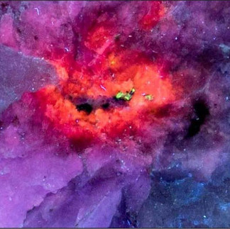
There is a second type of alteration of the sodalite. The second alteration is due to radiation damage. The alteration appears as a bright yellow-orange 1cm spot in the longwave UV image and a red-orange spot in the shortwave UV image. The radiation alteration occurred after the first alteration. In addition to changing the fluorescent response, the sodalite is no longer phosphorescent in the altered area. Pictures 8 through 11 show closeup pictures of the radiation alteration. At the center of the of the alteration are black grains of uraninite. The uraninite was emplaced with the dolomite. There are also grains of a shortwave UV green fluorescent uranium mineral along with the uraninite. Total radioactivity of the altered area is 265cpm compared to background of 24cpm on other parts of the specimen.
Picture 1 – white light image of brecciated sodalite.
Picture 2 – longwave UV image of the brecciated sodalite
Picture 3 – shortwave UV image of the brecciated sodalite
Picture 4 – afterglow after exposure to shortwave UV light
Picture 5 – white light image showing the tenebrescence
Picture 6 – longwave UV image showing the location of spectral measurements in Picture 7. Numbers correspond to numbers in Picture 7
Picture 7 – longwave UV fluorescent spectra of different sodalite types
Picture 8 – Closeup image of longwave UV fluorescence of radiation altered sodalite
Picture 9 -- Closeup image of shortwave UV fluorescence of radiation altered sodalite
Picture 10 – Closeup image of afterglow
Picture 11 – Closeup white light image of tenebrescence